Supercoiled DNA is far more dynamic than the “Watson-Crick” double helix
Researchers have imaged in unprecedented detail the three-dimensional structure of supercoiled DNA.

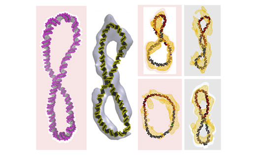
Various DNA shapes, including figure-8s, were imaged using a powerful microscopy technique by researchers at the Baylor College of Medicine in the US, and then examined using supercomputer simulations run at the University.
Improving our understanding of what DNA looks like when it is in the cell will help us to design better medicines, such as new antibiotics or more effective cancer chemotherapies.
Dr Sarah Harris from the School of Physics and Astronomy, who led the computer simulation research side of the study, said: “This is because the action of drug molecules relies on them recognising a specific molecular shape – much like a key fits a particular lock.”

The images show the structure of the DNA calculated with the supercomputer simulations (in colour) superimposed upon the cryo-electron tomography data (in white or yellow). You can see that the familiar double helix has been either simply bent into a circle or twisted into a figure-8.
The double helix shape has a firm place in the public's collective consciousness. It is referenced in popular culture and often features in art and design. But the shape of DNA isn’t always that simple.
Dr Harris said: “When Watson and Crick described the DNA double helix, they were looking at a tiny part of a real genome, only about one turn of the double helix. This is about 12 DNA ‘base pairs’, which are the building blocks of DNA that form the rungs of the helical ladder.
“Our study looks at DNA on a somewhat grander scale – several hundreds of base pairs – and even this relatively modest increase in size reveals a whole new richness in the behavior of the DNA molecule.”
Posted in: Research and innovation